13636351217

13636351217
聯系人:錢經理
電 話:13636351217
手 機:13636351217,13636351073
地 址:上海市松江臨港科技城漢橋文化科技園B座
郵 編:201615
傳 真:021-64881400
郵 箱:2881726255@qq.com
阿儀網商鋪:http://www.app17.com/c60514/
手機網站:m.shhyswkj.com
點擊次數:14603 發布時間:2020/12/30 14:01:28
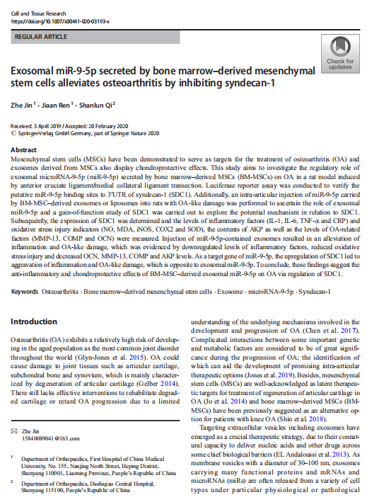
【文獻標題】Exosomal miR-9-5p secreted by bone marrow–derived mesenchymal stem cells alleviates osteoarthritis by inhibiting syndecan-1
【作者】Zhe Jin,Jiaan Ren,Shanlun Qi,et.al
【作者單位】中國醫科大學附屬第醫院(First Hospital of China Medical University)
【文獻中引用產品】
【關鍵詞】Osteoarthritis,Bone marrow–derived mesenchymal stem cells,Exosome,microRNA-9-5p,Syndecan-1
【DOI】https://doi.org/10.1007/s00441-020-03193-x
【影響因子(IF)】3.11
【出版期刊】《Cell and Tissue Research》
【產品原文引用】
Immunohistochemistry
The articular cartilage tissues were fixed in 4% paraformaldehyde for 24 h. Then, the tissues were dehydrated with gradient ethanol, paraffin-embedded and sliced into 5-μm sections. After being dewaxed, the tissue sections were dehydrated by gradient ethanol, immersed in 3% H2O2 for 10 min, followed by high-pressure antigen retrieval for 90 s. Afterwards, the sections were blocked with 100 μL 5% bovine serum albumin (BSA) solution at 37 °C for 30 min. The sections were then incubated with 100 μL rabbit anti SDC1 (1:500, ab128936, Abcam,Cambridge, MA, USA) overnight at 4 °C. The next day,the sections were incubated with biotin-labeled secondary antibody goat anti-rabbit (HY90046, Shanghai Hengyuan Biotechnology Co., Ltd., Shanghai, China) at 37 °C for 30 min. Then, the sections were incubated with streptavidin-peroxidase (Beijing Zhongshan Biotechnology Co., Ltd., Beijing, China) at 37 °C for 30 min and colored by diaminobenzidine (DAB) (Bioss Biotech, Beijing, China) at room temperature. Finally, the sections were counterstained by hematoxylin for 5 min,differentiated by 1% hydrochloric acid alcohol for 4 s and blued under running water for 20 min. The SDC1 positive cells were analyzed using Image-Pro Plus image analysis software (Media Cybernetics, Silver Springs,MD, USA). The brownish-yellow colored cells were considered as positive cells (Kelkar et al. 2017). Five highpower fields (× 200) were randomly selected from each section, with 100 cells counted in each field. The percentage of positive cells = the number of positive cells/the number of total cells and the percentage of positive cells > 10% was regarded as positive (+), < 10% as negative (−).The experiment was repeated three times independently.


完整版PDF文獻請咨詢在線客服或者電話聯系我司業務員!
更多公司福利請關注“恒遠生物”微信公眾號!

原創作者:上海恒遠生物技術發展有限公司